Phage characterization 噬菌体特征
The quality tests of the PVS also include a thorough phage characterization. ΦSB plaque morphology in PAO1 and PA4.6C showed uniform plaques with halos in 0.75% top layer LB agar (Supplementary Fig. S1A,B). The host range of ΦSB (Supplementary Table S1) displayed clear plaque formation spanning over 28.6% (14/49) of clinical Pseudomonas aeruginosa (PSA) strains isolated from sputum. Additionally, clear plaques were seen on 2/3 of non-pulmonary clinical PSA strains. Turbid plaques were observed for 17.3% (9/52) of all clinical PSA strains. Six clinical E. coli strains tested (negative controls) did not reveal plaques after spotting ΦSB.
PVS 的质量检测还包括对噬菌体的全面鉴定。PAO1和PA4.6C中的ΦSB菌斑形态在0.75%顶层LB琼脂中显示出带光晕的均匀菌斑(补充图S1A,B)。从痰中分离的临床铜绿假单胞菌(PSA)菌株中,28.6%(14/49)的 ΦSB 宿主范围(补充表 S1)显示出清晰的斑块形成。此外,2/3 的非肺部临床 PSA 菌株也出现了透明斑块。在所有临床 PSA 菌株中,17.3%(9/52)的菌株出现浑浊斑块。经测试的 6 株临床大肠杆菌(阴性对照)在点检 ΦSB 后未发现斑块。
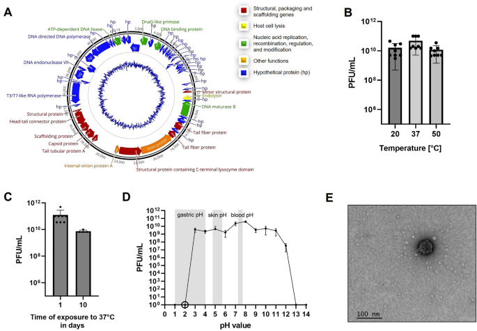
To study phage stability, ΦSB was stored in PBS supplemented with 1 M MgSO4 at 2e11 PFU/mL at 4 °C for four months, and separately for seven days in 0.9% saline at 1e9 PFU/mL at 4 °C without evidence of a significant decrease in viral titer (Supplementary Fig. S2). Thermal stability is presented in Fig. 2B,C. The effect of pH was studied for 24 h (Fig. 2D) with evidence that pH < 3 or pH > 11 decreased phage titer below detection limit (Fig. 2D), which was also observed for a 2-h exposure at pH 2 (Fig. 2D). Phage titers decreased 2–3.5 logs in serum and plasma at 37 °C (Supplementary Fig. S3). There were no differences between LDH in cell culture supernatants after phage incubation compared to controls (data not shown).TEM (Fig. 2E) indicated Autographiviridae morphology for ΦSB60.
为了研究噬菌体的稳定性,将ΦSB以2e11 PFU/mL的浓度在4 °C下保存在补充了1 M MgSO 4 的PBS中4个月,并分别以1e9 PFU/mL的浓度在4 °C下保存在0.9%生理盐水中7天,没有证据表明病毒滴度会显著下降(补充图S2)。热稳定性见图 2B、C。对 pH 值的影响进行了 24 小时的研究(图 2D),有证据表明,pH < 3 或 pH > 11 会使噬菌体滴度降至检测限以下(图 2D),在 pH 值为 2 时暴露 2 小时也观察到了这种情况(图 2D)。37 °C 时,血清和血浆中的噬菌体滴度下降了 2-3.5 个对数值(补充图 S3)。噬菌体培养后细胞培养上清液中的 LDH 与对照组相比没有差异(数据未显示)。

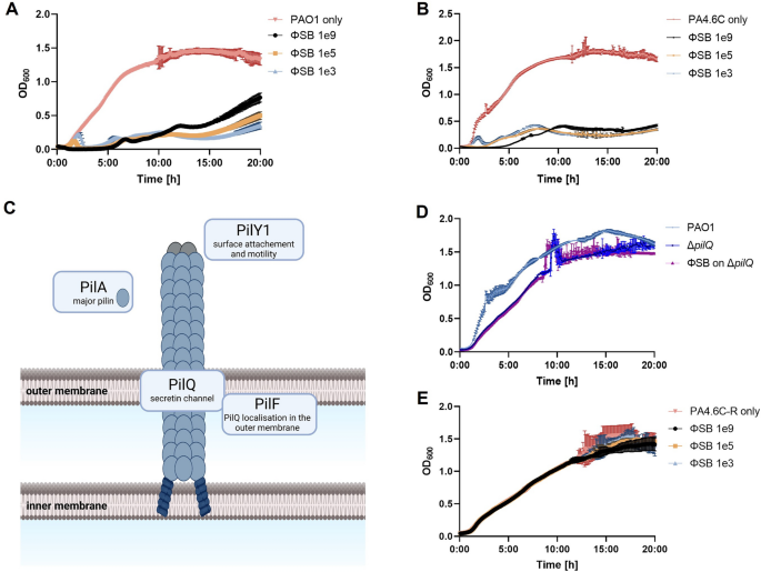
To study suppression of PSA growth, ΦSB was added to PSA cultures at a concentration of 1e9, 1e5, and 1e3 PFU/mL for 20 h. ΦSB decreased growth of PAO1 (Fig. 3A) and PA4.6C (Fig. 3B) compared to bacteria alone.
为研究 PSA 生长抑制情况,向 PSA 培养物中添加浓度为 1e9、1e5 和 1e3 PFU/mL 的 ΦSB 20 小时。与单独使用细菌相比,ΦSB 可减少 PAO1(图 3A)和 PA4.6C(图 3B)的生长。

Screening of transposon knockout strains revealed phage resistance in pilus-related knockout strains (ΔpilA, ΔpilY1, ΔpilQ, ΔpilF, Fig. 3C61,62,63,64, by spot assay. No phage amplification was observed within the incubation period, strongly suggesting that phage infection is impeded when a gene contributing to the construction of type-IV pili (TIVP) is knocked out. To examine this more closely, resistance towards the ΔpilQ-strain was confirmed by co-incubation of ΔpilQ-strain with ΦSB for 20 h (Fig. 3D). Efficiency of plating (EOP), which is the plating ability for a phage on the mutant strain relative to its plating ability on an ancestral phage sensitive host, could not be calculated because no visible plaques were produced on mutant strain ΔpilQ at 1e12 PFU/mL.
通过对转座子基因敲除菌株的筛选,发现与柔毛相关的基因敲除菌株(ΔpilA、ΔpilY1、ΔpilQ、ΔpilF,图 3C 61,62,63,64 )具有噬菌体抗性。通过定点检测。在孵育期内没有观察到噬菌体扩增,这有力地表明,当有助于构建第四型纤毛(TIVP)的基因被敲除时,噬菌体的感染会受到阻碍。为了更仔细地研究这一点,通过将 ΔpilQ 菌株与 ΦSB 共同培养 20 小时,证实了对ΔpilQ 菌株的抗性(图 3D)。由于在 1e12 PFU/mL 的条件下,突变菌株 ΔpilQ 上没有产生可见的斑块,因此无法计算噬菌体在突变菌株上的接种效率(EOP),即噬菌体在突变菌株上的接种能力与其在祖先噬菌体敏感宿主上的接种能力的比较。
Adsorption assays (Supplementary Fig. S4) showed evidence for ΦSB adsorption to host cells within 10 min and the one step growth curve (Supplementary Fig. S5) showed no discernable latent period, which prevented exact burst size calculation. Within 1 h and after initial MOI ~ 0.01, phage titer increased in the one step growth curve by an average of 3.77e2 ± 4.70e2 standard deviation.
吸附试验(补充图 S4)显示,ΦSB 在 10 分钟内吸附到宿主细胞上,一步生长曲线(补充图 S5)显示没有明显的潜伏期,因此无法准确计算猝灭规模。在初始 MOI ~ 0.01 后的 1 小时内,噬菌体滴度在一步生长曲线上平均增加了 3.77e2 ± 4.70e2 标准偏差。
To assess the potential capability to inhibit biofilm formation and to disrupt biofilms, crystal violet staining and CFU counts of biofilm assays revealed no viable bacteria in the biofilm formation assay and a significant reduction of pre-formed biofilm by both single phage treatments observed by crystal violet staining (p < 0.005, Supplementary Fig. S6A) and CFU (p < 0.005, Supplementary Fig. S6B).
为了评估噬菌体抑制生物膜形成和破坏生物膜的潜在能力,对生物膜进行了水晶紫染色和 CFU 计数,结果显示,在生物膜形成试验中没有存活细菌,而通过水晶紫染色(p < 0.005,补充图 S6A)和 CFU(p < 0.005,补充图 S6B)观察到,单一噬菌体处理可显著减少预先形成的生物膜。
